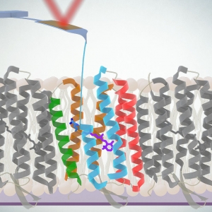
When it comes to drug development, membrane proteins play a crucial role, with about 50% of drugs targeting these molecules. Understanding the function of these membrane proteins, which connect to the membranes of cells, is important for designing the next line of powerful drugs. To do this, scientists study model proteins, such as bacteriorhodopsin (bR), which, when triggered by light, pump protons across the membrane of cells.
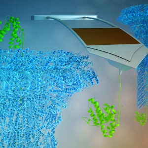
While bR has been studied for half a century, physicists have recently developed techniques to observe its folding mechanisms and energetics in the native environment of the cell’s lipid bilayer membrane. In a new study published by Proceedings of the National Academy of Sciences (PNAS), JILA and NIST Fellow Thomas Perkins and his team advanced these methods by combining atomic force microscopy (AFM), a conventional nanoscience measurement tool, with precisely timed light triggers to study the functionality of the protein function in real-time.

 and
and 


 lab")


