DNA imaging, ready in five minutes

New, fast technique produces clear images of DNA
AFM apex runs over a strand of DNA

A new technique from the Perkins group allows crisp, clear AFM images of DNA. The best part? It's ready in 5 minutes.

Image Credit
Steven Burrows and the Perkins Group/JILA

"I kind of think of it like a bowl of spaghetti noodles. If you drop them on the floor, they'd be kind of all globbed up and in weird shapes." - Patrick Heenan

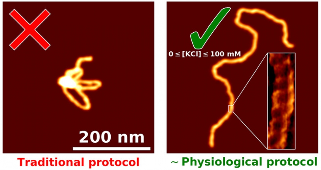
It’s tricky to get a good look at DNA when it won’t stick to your slide, or lay down in a straight strand.

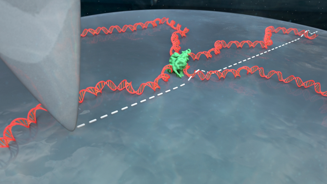
CU biochemist Tom Cech came to Tom Perkins’ atomic force microscopy lab with this very problem. His group was trying to understand how certain proteins interact with DNA. They knew their proteins interacted with DNA but they couldn’t get a good image of that interaction in liquid, DNA’s native environment.

“You have this nanoscale string in a ball shape that's undergoing thermal fluctuations, and then take this three-dimensional configuration, and squish it down really fast onto a surface,” Perkins explained. That squished DNA is often an uninterpretable blob.

“I kind of think of it like a bowl of spaghetti noodles. If you drop them on the floor, they'd be kind of all globbed up and in weird shapes,” explained Patrick Heenan, a graduate student in Perkins’ group.

The goal was to develop a process that would separate those globs into individual noodles. Heenan started working on this problem about two years ago, and he developed a technique to get your slide perfectly prepared to image a DNA sample. The best part? It’s fast – ready in just five minutes.

Tiny globs of DNA

DNA is about 2 nanometers wide – 100,000 times thinner than a sheet of paper. To get a good look at it, you need to put it on a slide that is truly flat. Mica, a light, soft silicate mineral, is a great candidate for a slide. All it takes to get a flat, thin sheet of mica is a piece of Scotch tape and some deft hands, Heenan explained.

“The beauty is it's super, super flat, so it is the perfect substrate,” Heenan said. “You can see all the atoms, and in some cases, you can see voids in the lattice.”

However, mica and DNA are both negatively charged; they repel each other. Scientists had tried changing the surface of the mica to make the DNA stick – soaking it for hours or days in various coatings and rinsing it with distilled water.

But the DNA still ends up in globs. When the apex of the atomic force microscope (AFM) tip runs over the DNA, the picture comes out blurry. Scientists can’t see how proteins or other molecules are interacting with the DNA. Many had given up on imaging DNA in liquid on bare mica, figuring it didn’t work or using coatings that made the images blurrier but still yielded globs. So Heenan started digging through the literature to see what others had tried.

“I read lots of papers, and I prepped hundreds of samples,” Heenan said. “It was a little bit like whacking through the weeds.”

Side-by-side comparison of traditional protocol versus Patrick Heenan's new method

Side-by-side comparison of traditional protocol versus Patrick Heenan's new method for imaging DNA with atomic force microscopy. The image on the right shows that the double helix of the DNA is clearly visible.

Image Credit
Perkins Group/JILA

Ready in five

It turned out soaking the mica for too long was part of the problem. Plus, the DNA wasn’t able to equilibrate on the surface, resulting in the squished ball shapes rather than nice, separate strands. “If you let it sit for a long time, you're actually losing some of the surface charge and impurities in the water are being sucked down into the surface,” Perkins explained.

Here’s how the improved process works. The mica is pre-soaked in a concentrated nickel-salt solution, then rinsed and dried. Then the DNA is bound to the mica with a solution of magnesium chloride and potassium chloride. Those conditions closely resemble the salts found naturally in cells, allowing the proteins and DNA to behave the way it normally would. Finally, the mica is rinsed with a nickel-chloride solution, which helps the DNA structure stick to the mica more tightly.

“It's kind of like the whole surface is uniformly sticky, so it can stick wherever it wants, and therefore it's easier for it to move around,” Heenan explained. “Everything is equally sticky.”

And it’s all ready for imaging in five minutes. Not only is it ready, but it delivers a clear picture – so clear that the double helix of the DNA is visible.

This development will help scientists study how DNA, protein and other molecules interact, as well as how DNA repairs itself.

“Patrick took on the challenge to image in liquid at biochemically-relevant conditions rather than in air, which, prior to Patrick's work, we and others would have assumed was impossible,” Perkins said. “What Patrick has developed is something that once you learn the eight steps, almost anybody can do it. It's really simple. It uses common salts, it takes five minutes, and you get higher signal-to-noise ratio, so basically, all sorts of good things.”

The study was published in ACS Nano on April 2, 2019, and was supported by the National Science Foundation Physics Frontier Grant and NIST.

Written by Rebecca Jacobson

Synopsis

Researchers at JILA have developed a fast, simple method to prepare samples that enhances DNA imaging. The results are so clear that the double-helix shape of DNA can be seen clearly.

Principal Investigators