Research overview

Homepage image.

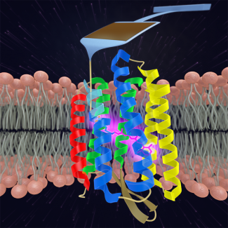
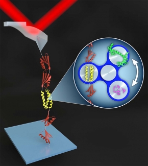
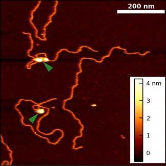
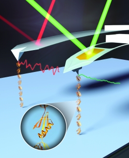
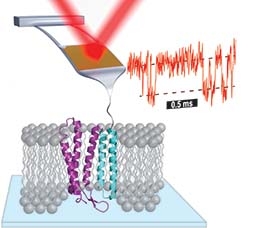
In the Perkins group, we develop and apply high-precision single-molecule techniques—atomic force microscopy (AFM) and optical traps—to address outstanding questions in a wide range of biological systems. Ongoing projects include elucidating the energetics and folding dynamics of membrane proteins embedded in their native lipid bilayer, characterizing the force needed to extract tubulin from a microtubule, probing the nanomechanics of myosin, and studying the motion of helicases along DNA. Our biological goals often motivate improvements in single-molecule techniques, such as developing focused-ion-beam-modified AFM cantilevers, an optical-trapping microscope with 1 Å stability in 3D, efficient site-specific conjugation strategies, and novel DNA- and protein-based constructs.

Research Areas

  • We are studying the energetics that stabilize membrane proteins by folding and unfolding individual molecules using AFM. By using cantilevers optimized for 1-µs resolution, we can measure the unfolding of individual molecules of the model membrane protein bacteriorhodopsin (bR) at 100-fold higher time resolution and 10-fold higher force precision than prior studies. This improvement in instrumental performance has allowed us to identify the energy barriers between states and to distinguish among obligatory, non-obligatory, and off-pathway intermediates.

  • Over the last 50 years, the search to understand the protein-folding process has blossomed into a large, interdisciplinary field. More recently, mechanobiology—the study of the mechanical forces exerted on molecules and cells—has also become an important and rapidly growing field. In both fields, the energy landscape provides the framework to describe the process of protein folding and how proteins respond to force. Local and global minima on this landscape represent intermediate and final states, respectively. In single molecule force spectroscopy (SMFS), regions of the energy landscape away from the global minimum are probed by perturbing a biomolecule with applied force, just as global denaturants such as urea or temperature are used in traditional biochemical assays.

  • We apply optical traps and AFM to yield mechanistic insight into how enzymes move along and bind to DNA as well as DNA’s fundamental mechanical properties. We developed an actively stabilized, optical-trapping microscope and applied it to study RecBCD, a helicase, at 1-bp resolution. More recently, we developed new techniques for AFM imaging of DNA in liquid and applied them to visualize the Polycomb repressive complex 2 (PRC2) bound to DNA. These new AFM measurements build on extensive prior studies of Polycomb group proteins by showing that PRC2 compacts DNA independent of its methyltransferase activity.

  • Single-molecule studies are a powerful means to study the dynamics and energetics of biological molecules and biomolecular complexes. These studies are limited by instrumentation resolution and assay efficiency. Historically, AFM was considered to have force resolution of ~5–20 pN and to suffer from significant instrumental drift. Over the last 15 years, we have substantially improved the precision, stability, and temporal resolution of bioAFM, culminating in studying membrane-protein unfolding with a 100-fold improvement in time resolution and a 10-fold improvement in force precision.