The Raschke group has created an ultrafast optical nanoscope based on a unique way of “nano” focusing the light to image the behavior of electrons on a thin gold film. The nanoscope is 1,000 times more powerful than conventional optical microscopes. It allows the researchers to investigate matter on its natural time and length scales, which are measured in femtoseconds and nanometers, respectively. This new technique may find application to studies of photosynthesis, solar cells, energy conversion and use, and other phenomena based on the transfer of electrons from molecule to molecule.
The nanoscope can see the motion of atoms and electrons at a speed that is one trillion times faster than the blink of an eye. This rate of image capture allowed the researchers to make a movie of the oscillations of electron clouds in real time and space! The researchers responsible for this amazing feat include graduate student Vasily Kravtsov, research associate Ronald Ulbricht, former research associate Joanna Atkin (University of North Carolina, Chapel Hill), and Fellow Markus Raschke.
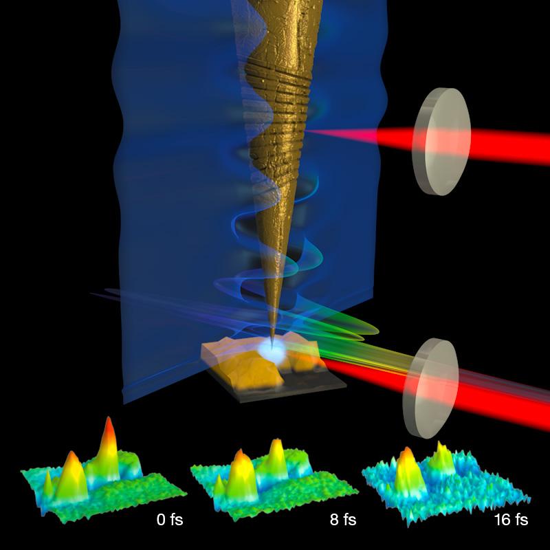
Their secret was figuring out how to nanofocus ultrashort light pulses on the tip of the scanning-probe nanoscope. The researchers accomplished this by generating plasmons on the nanometer-sized conical tip surface. In contrast to conventional light waves, plasmons are surface waves of light bound to the metallic tip shaft. They travel down to the tip of the cone. During their journey, they get so compressed that by the time a plasmon wave reaches the nanoscopic tip, its field strength is so large that it creates a rainbow of new colors of light. When the tip is then raster scanned [1] near the sample, a spatial image is created with individual movie frames that reflect the time delay of the excitation pulses.
“This work expands the reach of optical microscopes into the ultrafast and ultrasmall,” said Raschke. “Using this technique, researchers can image the elementary processes in materials ranging from battery electrodes to solar cells, helping to improve their efficiency and lifetime.”
The new technique of ultrafast optical nanoimaging will also help research groups to study charge and energy transport in soft matter, including biological materials.––Julie Phillips
[1] A raster scan is the rectangular pattern of image capture and reconstruction used in television