
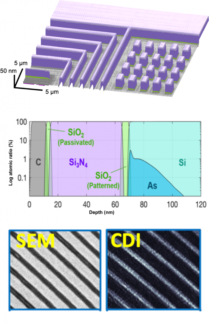
Figure. Coherent EUV beams can non-destructively image materials and buried structures, with chemical and compositional information, and diffraction-limited spatial resolution (~12 nm transverse, ~Å axial). (top and center) Coherent imaging of a semiconducting sample from imec, showing transverse and depth-resolved composition and doping. (bottom) First sub-wavelength imaging at short wavelengths.
Pushing short wavelength imaging to the fundamental limits
Although x-ray imaging has been explored for decades, and visible-wavelength microscopy for centuries, it is only recently that the spectral region in between―the extreme ultraviolet (EUV)―has been explored for imaging nanostructures and nanomaterials. With the practical implementation of coherent EUV light sources based on high harmonic generation (HHG), combined with coherent diffractive imaging (CDI), we have shown that EUV imaging has unique advantages. This is important because for synthesis and integration of a host of next-generation materials and nanostructures, new approaches are needed to non-destructively and routinely determine interfacial and layer structure as well as surface morphology, with sensitivity to dopant distributions and material composition
In recent research we demonstrated the first sub-wavelength imaging at short wavelengths using any light source, small or large.[6] This microscope achieved ~12 nm transverse, and ~Å axial resolution. We also developed the first full field dynamic imaging microscope, with 10fs temporal resolution. Finally, we developed the first phase-sensitive EUV imaging reflectometer. It combines the excellent phase stability of coherent high harmonic sources with the unique chemical- and phase- sensitivity of EUV reflectometry, and with state-of-the-art ptychography imaging algorithms. This tabletop microscope can non-destructively probe surface topography, layer thicknesses, and interface quality, as well as dopant concentrations and profiles.
Related Publications
-
M. Tanksalvala, C. Porter, Y. Esashi, G.Miley, N. Horiguchi, R. Karl, P. Johnsen, C. Bevis, N. Jenkins, B. Wang, X. Zhang, S. Cousin, D. Adams, M. Gerrity, H. Kapteyn, M. Murnane, “Non-Destructive, High-Resolution, Chemically Specific, 3D Nanostructure Characterization using Phase-Sensitive EUV Imaging Reflectometry,” submitted (2020).
-
R. Karl Jr., G. Mancini, J. Knobloch, T. Frazer, J. Hernandez-Charpak, B. Abad, D. Gardner, E. Shanblatt, M. Tanksalvala, C. Porter, C. Bevis, D. Adams, H. Kapteyn, M. Murnane, “Full-field imaging of thermal and acoustic dynamics in an individual nanostructure using tabletop high harmonic beams,” Science Advances 4, eaau4295 (2018). DOI: 10.1126/sciadv.aau4295
-
G. Mancini, R. Karl, E. Shanblatt, C. Bevis, D. Gardner, M. Tanksalvala, J. Russell, D. Adams, H. Kapteyn, J. Badding, T. Mallouk, M. Murnane, “Colloidal crystal order and structure revealed by tabletop extreme ultraviolet scattering and coherent diffractive imaging,” Optics Express 26(9) 11393–11406 (2018). DOI:10.1364/OE.26.011393
-
C. Bevis, R. Karl Jr., J. Reichanadter, D. Gardner, C. Porter,E. Shanblatt, M. Tanksalvala, G. Mancini, H. Kapteyn, M. Murnane, D. Adams, “Multiple beam ptychography for large field-of-view, high throughput, quantitative phase contrast imaging,” Ultramicroscopy 184, 164–171 (2018). DOI: 10.1016/j.ultramic.2017.08.018
-
C. Porter, M. Tanksalvala, M. Gerrity, G. Miley, X. Zhang, C. Bevis, E. Shanblatt, R. Karl, M. Murnane, D. Adams, H. Kapteyn, “General-purpose, wide field-of-view reflection imaging with a tabletop 13nm light source,” Optica 4(12) 1552–1557 (2017). DOI: 10.1364/OPTICA.4.001552
-
D. Gardner, M. Tanksalvala, E. Shanblatt, X. Zhang, B. Galloway, C. Porter, R. Karl Jr., C. Bevis, D. Adams, H. Kapteyn, M. Murnane, G. Mancini, “Subwavelength coherent imaging of periodic samples using a 13.5 nm tabletop high-harmonic light source,” Nature Photonics 11, 259 (2017). DOI:10.1038/nphoton.2017.33
-
E. Shanblatt, C. Porter, D. Gardner, G. Mancini, R. Karl Jr., M. Tanksalvala, C. Bevis, V. Vartanian, H. Kapteyn, D. Adams, M. Murnane, “Quantitative Chemically Specific Coherent Diffractive Imaging of Reactions at Buried Interfaces with Few Nanometer Precision,” Nano Letters 16 (9), 5444–5450 (2016). DOI: 10.1021/acs.nanolett.6b01864
-
B. Zhang, D. F. Gardner, M. D. Seaberg, E. R. Shanblatt, Henry C. Kapteyn, Margaret M. Murnane, D. E. Adams, “High contrast 3D imaging of surfaces near the wavelength limit using tabletop EUV ptychography,” Ultramicroscopy 158, 98–104 (2015). https://doi.org/10.1016/j.ultramic.2015.07.006 Featured on Cover.
-
J. Miao, T. Ishikawa, I. K. Robinson, M. M. Murnane, “Beyond crystallography: Diffractive imaging using coherent X-ray light sources,” Science 348, 530 (2015). DOI: 10.1126/science.aaa1394 Featured on cover of Science.
-
M. D. Seaberg, B. Zhang, D. F. Gardner, E. R. Shanblatt, M.M. Murnane, H. C. Kapteyn, D.E. Adams, “Tabletop nanometer extreme ultraviolet imaging in an extended reflection mode using coherent Fresnel ptychography,” Optica 1, 39 (2014). https://doi.org/10.1364/OPTICA.1.000039
-
B. Zhang, M. Seaberg, J. Shaw, D. Gardner, D. Adams, M. Murnane, H. Kapteyn, “Full field tabletop EUV coherent diffractive imaging in a transmission geometry,” Optics Express 21, 21970 (2013). https://doi.org/10.1364/OE.21.021970).
-
R.L. Sandberg, D. A. Raymondson, C. La-o-vorakiat, A. Paul, K. S. Raines, J. Miao, M. M. Murnane, H. C. Kapteyn, W. F. Schlotter, “Tabletop soft x-ray Fourier transform holography with 50 nm resolution,” Optics Letters 34, 1618 (2009). https://doi.org/10.1364/OL.34.001618
-
R. L. Sandberg, A. Paul, D. A. Raymondson, S. Hädrich, D. M. Gaudiosi, J. Holtsnider, R. Tobey, O. Cohen, M. M. Murnane, H. C. Kapteyn, C. Song, Ji. Miao, Y. Liu, F. Salmassi, “Lensless diffractive imaging using tabletop coherent high-harmonic soft-X-ray beams,” Physical Review Letters 99, 098103 (2007). DOI: 10.1103/PhysRevLett.99.098103