


 lab")


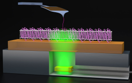
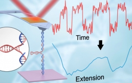
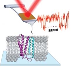
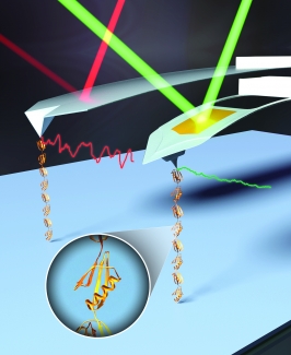
We are studying the energetics that stabilize membrane proteins by folding and unfolding individual molecules using AFM. By using cantilevers optimized for 1-µs resolution, we can measure the unfolding of individual molecules of the model membrane protein bacteriorhodopsin (bR) at 100-fold higher time resolution and 10-fold higher force precision than prior studies. This improvement in instrumental performance has allowed us to identify the energy barriers between states and to distinguish among obligatory, non-obligatory, and off-pathway intermediates.
About the Perkins Group
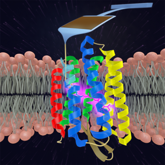
In the Perkins group, we develop and apply high-precision single-molecule techniques—atomic force microscopy (AFM) and optical traps—to address outstanding questions in a wide range of biological systems. Ongoing projects include elucidating the energetics and folding dynamics of membrane proteins embedded in their native lipid bilayer, characterizing the force needed to extract tubulin from a microtubule, probing the nanomechanics of myosin, and studying the motion of helicases along DNA. Our biological goals often motivate improvements in single-molecule techniques, such as developing focused-ion-beam-modified AFM cantilevers, an optical-trapping microscope with 1 Å stability in 3D, efficient site-specific conjugation strategies, and novel DNA- and protein-based constructs.
Stories About Our Research
Probing Proton Pumping: New Findings on Protein Folding in bacteriorhodopsin (bR)
When it comes to drug development, membrane proteins play a crucial role, with about 50% of drugs targeting these molecules. Understanding the function of these membrane proteins, which connect to the membranes of cells, is important…
Read MoreThe Forces involved in Folding Proteins
In a new paper, JILA physicist Thomas Perkins collaborated with CU Biochemistry Prof. Marcello Sousa to dissect the mechanisms of how certain bacteria become more virulent. The research brings together the Perkins lab expertise in…
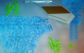
Read MoreGrabbing Proteins by the Tail
"Unraveling" cell membrane proteins could help us understand how to build better drugs and treatments for disease.
Read More
DNA imaging, ready in five minutes
It's tough to get tightly-wound balls of DNA to lay down flat and straighten out to get their picture taken. A new technique from the Perkins group gets a crisp, clear picture in just five minutes.
Read MorePulling apart HIV
JILA researchers have demonstrated a much easier, faster and more precise way to understand the structure and function of the HIV RNA molecule, especially the HIV RNA hairpin. Furthermore, the techniques developed for this research…
Read MorePrecision Biomechanics
The Perkins group has made dramatic advances in the use of Atomic Force Microscopes (AFMs) to study large single biomolecules, such as proteins and nucleic acids (DNA, RNA), that are important for life. After previously improving AFM…
Read More
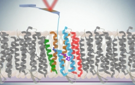
Vision Quest
The Perkins group continues to extend the performance of its unique Atomic Force Microscope (AFM) technology, revealing for the first time a dozen new short-lived intermediate states in the folding and unfolding of a membrane protein…
Read MoreThe Land of Enhancement: AFM Spectroscopy
The Perkins Group has demonstrated a 50-to-100 times improvement in the time resolution for studying the details of protein folding and unfolding on a commercial Atomic Force Microscope (AFM). This enhanced real time probing of protein…
Read MoreThe Measure of Small Things
Fellow Tom Perkins’ group is significantly closer to realizing its long-standing dream of using atomic force microscopy (AFM) to study how membrane proteins fold and unfold. Historically, scientists have used AFM to measure the…
Read More
bR Phone Home
The groups of Fellow Adjoint Markus Raschke and Fellow Tom Perkins joined forces recently to shine light onto a bacterial membrane protein called bacteriorhodopsin (bR). They used a new infrared (IR) light imaging system with a spatial…
Read MoreGoing for the Gold
Gold glitters because it is highly reflective, a quality once considered important for precision measurements made with gold-coated probes in atomic force microscopy (AFM). In reality, the usual gold coating on AFM probes is a major…
Read MoreUpending Conventional Wisdom
In science, it can be fun and interesting to upend conventional wisdom. A good example is what just happened to a widely accepted explanation for overstretching of double-stranded DNA (dsDNA). Overstretching occurs suddenly when…
Read More
The Guiding Light
Atomic force microscopy (AFM) just got a whole lot more efficient for studying proteins and other biomolecules. Graduate student Allison Churnside, former research associate Gavin King, and Fellow Tom Perkins recently used a laser to…
Read MoreHow to Marry a Microscope
The most important step for a microscope wanting to marry another microscope is finding the right partner. A professional matchmaker, such as the Perkins lab, might be just the ticket. The group recently presided over the nuptials of…
Read MoreDNA: Force of Nature
The Perkins group is helping to develop DNA as a force standard for the nano world. Polymers of DNA act like springs, and DNA's elasticity may one day provide a force standard from 0.1–10 piconewtons (pN). One pN is the force exerted…
Read More
Gold Fever
Life can be challenging on the biophysics research frontier. Consider gold nanoparticles as a research tool, for example. Gold is ductile and malleable as well as being a good conductor of heat and electricity. Its unique chemistry…
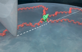
Read MoreSightseeing along a DNA Strand
Lora Nugent-Glandorf and Tom Perkins have come up with an optical trap motion detector that can "see" protein motors moving one base at a time along a DNA helix. For some time scientists have been able to make optical traps that can…
Read More
Research Highlights
Probing Proton Pumping: New Findings on Protein Folding in bacteriorhodopsin (bR)
When it comes to drug development, membrane proteins play a crucial role, with about 50% of drugs targeting these molecules. Understanding the function of these membrane proteins, which connect to the membranes of cells, is important…
Read More
The Forces involved in Folding Proteins
In a new paper, JILA physicist Thomas Perkins collaborated with CU Biochemistry Prof. Marcello Sousa to dissect the mechanisms of how certain bacteria become more virulent. The research brings together the Perkins lab expertise in…
Read More
Grabbing Proteins by the Tail
"Unraveling" cell membrane proteins could help us understand how to build better drugs and treatments for disease.
Read More
DNA imaging, ready in five minutes
It's tough to get tightly-wound balls of DNA to lay down flat and straighten out to get their picture taken. A new technique from the Perkins group gets a crisp, clear picture in just five minutes.
Read More
Pulling apart HIV
JILA researchers have demonstrated a much easier, faster and more precise way to understand the structure and function of the HIV RNA molecule, especially the HIV RNA hairpin. Furthermore, the techniques developed for this research…
Read More
Precision Biomechanics
The Perkins group has made dramatic advances in the use of Atomic Force Microscopes (AFMs) to study large single biomolecules, such as proteins and nucleic acids (DNA, RNA), that are important for life. After previously improving AFM…
Read More
Vision Quest
The Perkins group continues to extend the performance of its unique Atomic Force Microscope (AFM) technology, revealing for the first time a dozen new short-lived intermediate states in the folding and unfolding of a membrane protein…
Read More
The Land of Enhancement: AFM Spectroscopy
The Perkins Group has demonstrated a 50-to-100 times improvement in the time resolution for studying the details of protein folding and unfolding on a commercial Atomic Force Microscope (AFM). This enhanced real time probing of protein…
Read More
The Measure of Small Things
Fellow Tom Perkins’ group is significantly closer to realizing its long-standing dream of using atomic force microscopy (AFM) to study how membrane proteins fold and unfold. Historically, scientists have used AFM to measure the…
Read More
bR Phone Home
The groups of Fellow Adjoint Markus Raschke and Fellow Tom Perkins joined forces recently to shine light onto a bacterial membrane protein called bacteriorhodopsin (bR). They used a new infrared (IR) light imaging system with a spatial…
Read More
Going for the Gold
Gold glitters because it is highly reflective, a quality once considered important for precision measurements made with gold-coated probes in atomic force microscopy (AFM). In reality, the usual gold coating on AFM probes is a major…
Read More
Upending Conventional Wisdom
In science, it can be fun and interesting to upend conventional wisdom. A good example is what just happened to a widely accepted explanation for overstretching of double-stranded DNA (dsDNA). Overstretching occurs suddenly when…
Read More
The Guiding Light
Atomic force microscopy (AFM) just got a whole lot more efficient for studying proteins and other biomolecules. Graduate student Allison Churnside, former research associate Gavin King, and Fellow Tom Perkins recently used a laser to…
Read More
How to Marry a Microscope
The most important step for a microscope wanting to marry another microscope is finding the right partner. A professional matchmaker, such as the Perkins lab, might be just the ticket. The group recently presided over the nuptials of…
Read More
DNA: Force of Nature
The Perkins group is helping to develop DNA as a force standard for the nano world. Polymers of DNA act like springs, and DNA's elasticity may one day provide a force standard from 0.1–10 piconewtons (pN). One pN is the force exerted…
Read More
Gold Fever
Life can be challenging on the biophysics research frontier. Consider gold nanoparticles as a research tool, for example. Gold is ductile and malleable as well as being a good conductor of heat and electricity. Its unique chemistry…
Read More
Sightseeing along a DNA Strand
Lora Nugent-Glandorf and Tom Perkins have come up with an optical trap motion detector that can "see" protein motors moving one base at a time along a DNA helix. For some time scientists have been able to make optical traps that can…
Read More
Research Areas
Over the last 50 years, the search to understand the protein-folding process has blossomed into a large, interdisciplinary field. More recently, mechanobiology—the study of the mechanical forces exerted on molecules and cells—has also become an important and rapidly growing field. In both fields, the energy landscape provides the framework to describe the process of protein folding and how proteins respond to force. Local and global minima on this landscape represent intermediate and final states, respectively. In single molecule force spectroscopy (SMFS), regions of the energy landscape away from the global minimum are probed by perturbing a biomolecule with applied force, just as global denaturants such as urea or temperature are used in traditional biochemical assays.
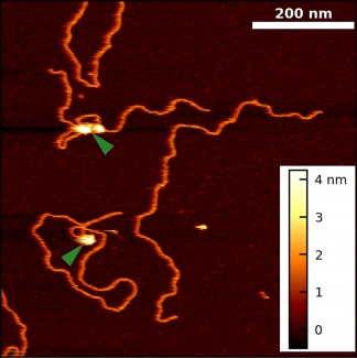
We apply optical traps and AFM to yield mechanistic insight into how enzymes move along and bind to DNA as well as DNA’s fundamental mechanical properties. We developed an actively stabilized, optical-trapping microscope and applied it to study RecBCD, a helicase, at 1-bp resolution. More recently, we developed new techniques for AFM imaging of DNA in liquid and applied them to visualize the Polycomb repressive complex 2 (PRC2) bound to DNA. These new AFM measurements build on extensive prior studies of Polycomb group proteins by showing that PRC2 compacts DNA independent of its methyltransferase activity.
Single-molecule studies are a powerful means to study the dynamics and energetics of biological molecules and biomolecular complexes. These studies are limited by instrumentation resolution and assay efficiency. Historically, AFM was considered to have force resolution of ~5–20 pN and to suffer from significant instrumental drift. Over the last 15 years, we have substantially improved the precision, stability, and temporal resolution of bioAFM, culminating in studying membrane-protein unfolding with a 100-fold improvement in time resolution and a 10-fold improvement in force precision.
In the Spotlight


JILA Address
 We are located at JILA: A joint institute of NIST and the University of Colorado Boulder.
Map | JILA Phone: 303-492-7789 | Address: 440 UCB, Boulder, CO 80309