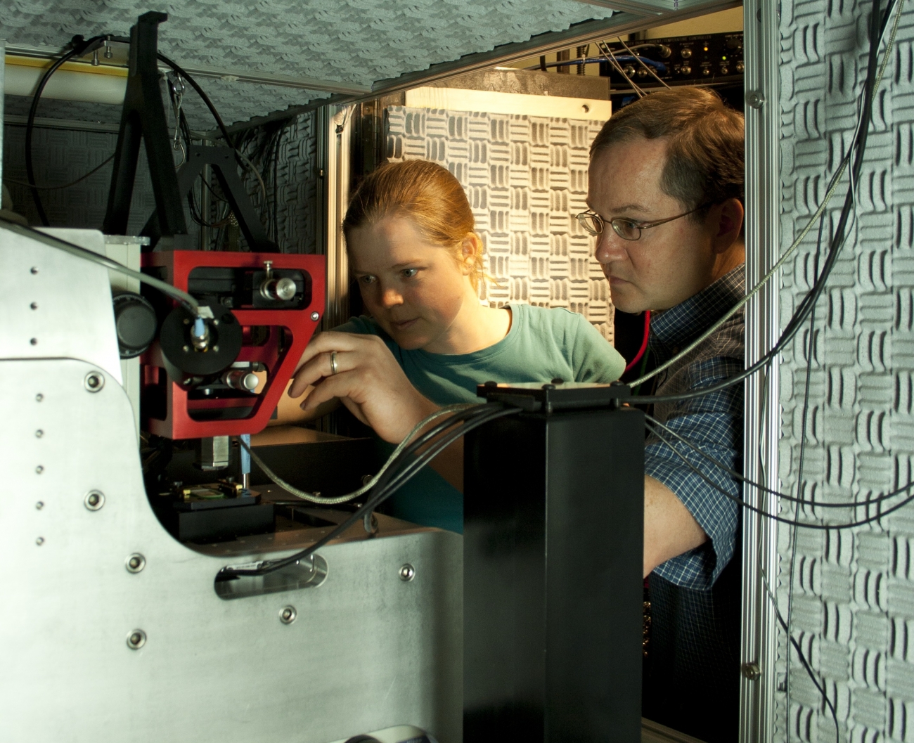
Atomic force microscopy (AFM) just got a whole lot more efficient for studying proteins and other biomolecules. Graduate student Allison Churnside, former research associate Gavin King, and Fellow Tom Perkins recently used a laser to detect the position of sparsely distributed biomolecules on a glass cover slip. Since the same laser is also used to locate the AFM tip, it is now possible to align the microscope tip and sample with a precision of 40 nm, before the AFM tip even touches the sample. The researchers say that the new sample detection scheme solves the “needle in a haystack” problem of nanoscale microscopy.
Until now, scientists employing AFM to study nanostructures had to use a kind of brute-force method to locate a single sample on a microscope substrate more than a million times bigger than a target biomolecule. The brute-force method involves rapidly scanning back and forth across the stage until the tip encounters something interesting. There are several problems with this method: (1) Since an AFM tip is only a few atoms wide, it is delicate and easy to break. (2) It’s easy to contaminate an AFM tip with unwanted atoms or molecules encountered during the scan, and AFM tips can form chemical bonds with both contaminants and biomolecules. Such chemical bonds could be a big advantage in studies of individual membrane proteins, for example, but only if the risk of tip contamination is low. (3) Biological samples, such as proteins or membranes, can be damaged by uncontrolled collisions with an AFM tip. A squished sample defeats the purpose of investigating intact biological nanostructures.
The new laser detection system protects both the AFM tip from physical damage and contamination as well as the sample from harm. Plus, it’s much more efficient. “From a practical perspective, instead of Alison starting to do real science at 4 p.m. after she’s spent most of a day looking for a good sample, she can start doing science at 10 a.m.,“ Perkins says. From here on, Perkins expects the use of AFM to be less frustrating and more likely to lead to interesting scientific results. For example, Churnside recently employed a focused laser beam to optically detect a purple membrane patch, which she then studied in more detail using AFM.
“Now we can use our new optical method to find a structure that looks like what we want to study, then drop the AFM tip and land “dead on” the exact spot we’re seeing,” Perkins says. As the new technique becomes more refined, he expects to be able to “see” smaller and smaller things. Currently, the lateral resolution of optical images is ~200 nm, a resolution that is set by the diffraction limit for optical microscopy. Nevertheless, optical images can “resolve” patches of purple membrane that are only 6 nm tall. The enhanced height resolution is due to the ability to distinguish the average height of a patch rather than having to distinguish two closely spaced objects, as must occur in lateral resolution. The Perkins group expects to be able to refine the vertical resolution down to ±1 nm or less. - Julie Phillips